Tips on Medication Management: Finding The Top and Bottom of The Therapeutic Window
January 28, 2008Solving Crazy Meetings: “Six Thinking Hats” Will Save the Day
January 31, 2008Brain, SPECT and Celiac
Brain changes do appear on SPECT imaging with bowel immune dysfunction: think gluten sensitivity and celiac. Seeing real pathological evidence, bottom to top, does help with believing. Just a few months ago we reported at CorePsych Blog the interesting findings, reported in the Annals of Internal Medicine, of all places, on SPECT brain imaging, celiac and a clearing of schizophrenic symptoms when the gluten sensitivity was effectively treated. Schizophrenia cured by eating a proper diet… and reported in an Internal Medicine journal? Interesting how others are seeing evidence for what some of us in mental health are missing.
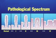
Gluten Villi in Stages of Deterioration
This pathology slide set shows what celiac and early gluten sensitivity look like on the bowel level from the Columbia University Celiac Disease Center:
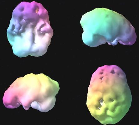
Just below: a typical brain image we often see using SPECT imaging for individuals with immune dysregulation. So many articles are coming up regarding brain imaging with SPECT with celiac I thought you would like to see the actual brain level findings that we see in our office.
The Immune Brain
Here you can easily see the temporal lobe and prefrontal hypoperfusion, so often referenced in the literature, that lead us to always chase down possible immune dysfunction/bowel/celiac issues as reported in the article below. -Yes, a picture is worth a thousand words…
Another article [ref PubMed] supports additional SPECT imaging evidence in celiac disease:
Frontal cortical perfusion abnormalities related to gluten intake and associated autoimmune disease in adult coeliac disease: 99mTc-ECD brain SPECT study. Usai P et al. Dig Liver Dis. 2004; 36(8): 513-8
See the conclusions below from that article:
RESULTS: Twenty-four out of 34 patients (71%) showed brain single-photon emission computed tomography abnormalities confirmed by abnormal regional asymmetry index (>5%; range 5.8-18.5%). Topographic comparison of the brain areas showed that the more significant abnormalities were localised in frontal regions, and were significantly different from controls only in coeliac disease patients on unrestricted diet. The prevalence of single-photon emission computed tomography abnormalities was similar in coeliac disease patients with (74%) and without (69%) associated autoimmune disease.
CONCLUSIONS: Abnormalities of brain perfusion seem common in coeliac disease. This phenomenon is similar to that previously described in other autoimmune diseases, but does not appear to be related to associated autoimmunity and, at least in the frontal region, may be improved by a gluten-free diet.
These interesting articles reveal the growing interface between brain and body evidence in immune dysfunction. It's interesting to bring these brain studies together with real office laboratory findings that confirm the pathophysiology on a gut cellular level.
Of further interest: SPECT is demonstrating changes in mold neurotoxin, neuroimmune conditions as well – more on mold findings in a later post.
cp
Dr Charles Parker
CoreBrain Journal Podcast Updates: http://corebrainjournal.com
CorePsych Inquires: Brief Note: This Link
YouTube: Connect & Subscribe For Updates: This Link
Complimentary & New: 23 pg Special Report: Predictable Solutions For ADHD Medications
International Book Link: New ADHD Medication Rules: http://geni.us/adhd
Related articles by Zemanta
- This is your brain on wheat II (heartscanblog.blogspot.com)
- Celiac Disease and The Gluten Free Diet (nutrition.suite101.com)
- Cereal Killers: Celiac Disease and Gluten-Free A To Z Reveals New Insights into the Nature of Celiac Disease and Gluten Sensitivity (prweb.com)
- Toxic trio identified as the basis of celiac disease (eurekalert.org)
- Celiac disease – sprue – All Information (umm.edu)
- Celiac disease – nutritional considerations – All Information (umm.edu)


4 Comments
[…] many of those images derived from immunity matters – not brain injury. That hypoperfusion looked like brain injury in the SPECT images – but without brain injury in the patient’s history the discovery imperative changed to […]
Celiacbrain is like a grain, that it why they look the same. Maybe proper exercising will help improve hippocampus grow.
Jackie,
Two very interesting questions, both relevant to every-day office practice:
Regarding testing: Having discussed this issue with a variety of my colleagues both traditional GI docs [many of whom remain locked on/searching only for “celiac” diagnostic findings through LabCorp and biopsy] and researchers like Neuroimmunologist Dr Vojdani [referenced in several previous posts as considerably interested in multiple immune dysregulations with subsequent brain/neurological pathology] the testing debate continues vigorously offline.
My current solution, argued against by some on both sides of the traditional/functional fence, is fecal testing [more easily accomplished with some folks] with Enterolabs.com [Dr Fine], and blood testing with Neuroscience Labs [Dr Vojdani] for a variety of immune dysregulation issues.
And the other question regarding SPECT and enzymes… interesting, and at this moment I am unfamiliar with any such brain studies. The use of enzymes, as a means of coping with gluten sensitivity, is still controversial as some ardently insist that the only way is total abstinence and others indicate they have had some success with enzymes.
My colleague, Dr Tom OBryan, who lectures nationally on all of these matters will be on a national tour soon this Spring and I will post here at CorePsychBlog on his speaking dates and locations.
A final thought, as your example is so frequently the office presentation – clear metabolic symptoms and refractory to conventional interventions – I would stick with your regular psych interventions, discuss the bio/complexity with your doc, read as much as you can starting with gluten sensitivity and go for the percentage improvement in diet.
Remember: Wheat is only one potential contributor to neuroimmune problems. Milk, soy and a variety of other foods can significantly create problems, as can mold and even aluminum from cans!
The good news is: all of these potential allergens can be tested for – and while a *grand consensus* may not exist at this moment on *how,* I have seen considerable progress by simply looking, measuring and pursuing this immunologic path of inquiry.
Thanks,
Chuck
Dr. Parker,
Thanks for this great blog!
When we had my daughter (adhd & ld) tested 10 years ago, she was negative for celiac disease. My two questions are:
1) is there currently a validated mainstream-established test for gluten intolerance that is not classic celiac? and
2) have there been any spect studies done that would indicate that the enzyme supplements on the market can counteract the brain abnormalities (similar to the way a gluten-free diet seems to?)
I ask this last question because my daughter is a teenager, and highly unlikely to stick consistently to ANY restrictive diet, at least for several years to come! However, clues that there may be something going on for her in this arena include: sensitivity to adhd meds (for example, she seems to metabolize lower doses of Vyvanse very quickly, but higher doses make her over-focused and angry), persistent eczema, and rhinitis (docs can’t seem to agree if it’s non-allergic or an allergy to something they just haven’t found yet!).
I know you’re going to ask me about bowel symptoms, but I’m really not sure — except to say that as a kid she leaned to the more infrequent/constipated side…
I look forward to your insights on this.